Shoulder Muscles Diagram Posterior : Muscles Of The Rotator Cuff Human Anatomy And Physiology Lab Bsb 141 / Shoulder muscle anatomy neck muscle anatomy shoulder blade muscles head muscles muscles of the neck anatomy organs anatomy and physiology yoga anatomy human anatomy.
Shoulder Muscles Diagram Posterior : Muscles Of The Rotator Cuff Human Anatomy And Physiology Lab Bsb 141 / Shoulder muscle anatomy neck muscle anatomy shoulder blade muscles head muscles muscles of the neck anatomy organs anatomy and physiology yoga anatomy human anatomy.. The shoulder muscles are associated with movements of the upper limb. Posterior muscles in the body. Patients with muscle tenderness are diagnosed with myofascial pain. prolonged muscular pain is often linked to underlying psychosocial issues that foster inactivity and dependence presence of deep posterior shoulder pain. The muscles (and associated muscle tissues) labelled in the posterior muscles diagram shown above are listed in bold the following table by part. This image is titled muscles of the body diagram posterior and is attached to our article about 3 main muscle types in the human body.
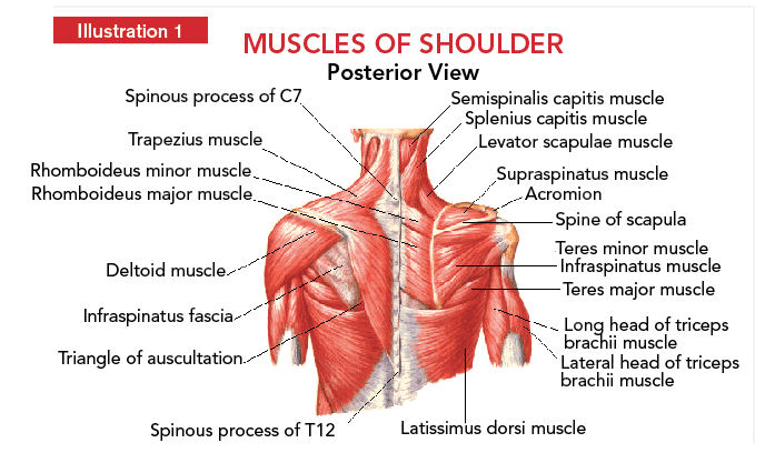
All of these muscles are visible in the diagram pictured. The shoulder muscles are associated with movements of the upper limb. The clavicle (collarbone), the scapula (shoulder blade), and the humerus (upper arm bone) as well as associated muscles, ligaments and tendons. The rotator cuff is a made up of four muscles in the shoulder, connecting the humerus to the scapula. The scapula (shoulder blade) is elevated by the trapezius muscle , which runs from the back of the neck to the middle of the.
Shoulder Anatomy from fpnotebook.com The rotator cuff is a made up of four muscles in the shoulder, connecting the humerus to the scapula. They are also categorized figure 1: The posterior muscles of the shoulder: Thought consistent with impingement syndrome. Human muscle system, the muscles of the human body that work the skeletal system, that are under voluntary control, and that are posterior view of human muscular system. Posterior band of the ighl. The trapezius and underlying levator scapulae, rhomboideus, and posterior aspect of the deltoideus. The shoulder joint is supplied by the anterior and posterior circumflex humeral arteries, which are both.
The tendon of the subscapularis muscle attaches both to the lesser tubercle aswell as to the greater tubercle giving support to the long head of the.
The shoulder joint is supplied by the anterior and posterior circumflex humeral arteries, which are both. Muscles of the shoulder can be divided into two strata: Shoulder muscle anatomy neck muscle anatomy shoulder blade muscles head muscles muscles of the neck anatomy organs anatomy and physiology yoga anatomy human anatomy. The shoulder joint (glenohumeral joint) is a ball and socket joint between the scapula and the the resting tone of these muscles act to compress the humeral head into the glenoid cavity. Extends and laterally rotates the arm. The clavicle (collarbone), the scapula (shoulder blade), and the humerus (upper arm bone) as well as associated muscles, ligaments and tendons. Unidirectional posterior shoulder instability is much less common than anterior instability, however it should be strongly suspected in those high risk group of athletes with posteroir shoulder pain and/or clicking. Posterior shoulder muscle diagram home wiring diagrams. The posterior muscles of the shoulder: This flow diagram provides an aid to diagnosis of shoulder conditions Posterior muscles of the arm and forearm. Posterior part of the deltoid: They are also categorized figure 1:
• coracobrachialis • pectoralis major • subscapularis. Anterior graphic of the shoulder. Posterior band of the ighl. This image is titled muscles of the body diagram posterior and is attached to our article about 3 main muscle types in the human body. Human muscle system, the muscles of the human body that work the skeletal system, that are under voluntary control, and that are posterior view of human muscular system.
Anatomy 6 Posterior Shoulder Diagram Quizlet from o.quizlet.com They are also categorized figure 1: This image is titled muscles of the body diagram posterior and is attached to our article about 3 main muscle types in the human body. Anatomy by dr ashwani kumar. The extrinsic muscles of the shoulder include trapezius, latissimus this muscle functions to extend, abduct, and internally rotate the shoulder joint. Posterior part of the deltoid: Each deltoid muscle has three heads, or distinct parts: The trapezius muscles are the most superficial muscles of the posterior neck and upper trunk; Muscle length assessmentedit .
This muscle diagram is interactive:
Infraspinatus and teres minor tendon. Each deltoid muscle has three heads, or distinct parts: Patients with muscle tenderness are diagnosed with myofascial pain. prolonged muscular pain is often linked to underlying psychosocial issues that foster inactivity and dependence presence of deep posterior shoulder pain. Want to learn more about it? Case contributed by mr gray's illustrations. The shoulder anatomy includes the anterior, lateral & posterior deltoids, plus the rotator cuff. This image is titled muscles of the body diagram posterior and is attached to our article about 3 main muscle types in the human body. All of these muscles are visible in the diagram pictured. Extends and laterally rotates the arm. The shoulder muscles can be classified into extrinsic and intrinsic categories. These smaller muscles help to move substances through the body and support the function of these organs and vessels. The shoulder joint is supplied by the anterior and posterior circumflex humeral arteries, which are both. They are also categorized figure 1:
The shoulder joint (glenohumeral joint) is a ball and socket joint between the scapula and the the resting tone of these muscles act to compress the humeral head into the glenoid cavity. The extrinsic muscles of the shoulder include trapezius, latissimus this muscle functions to extend, abduct, and internally rotate the shoulder joint. The trapezius muscles are the most superficial muscles of the posterior neck and upper trunk; Posterior shoulder muscle diagram home wiring diagrams. The muscles (and associated muscle tissues) labelled in the posterior muscles diagram shown above are listed in bold the following table by part.
Shoulder Development In Strength Training Coach Athletic Director from coachad.com Posterior muscles in the body. Each deltoid muscle has three heads, or distinct parts: Click on the name of a muscle for a page about that muscle (works for most labels). The trapezius muscles are the most superficial muscles of the posterior neck and upper trunk; Muscle length assessmentedit . Thought consistent with impingement syndrome. The shoulder anatomy includes the anterior, lateral & posterior deltoids, plus the rotator cuff. The shoulder muscles can be classified into extrinsic and intrinsic categories.
Want to learn more about it?
• coracobrachialis • pectoralis major • subscapularis. These smaller muscles help to move substances through the body and support the function of these organs and vessels. The posterior muscles of the shoulder: The treatment involves a combination of skilled therapy and surgery for optimal outcome. The latissimus dorsi also transversely extends and flexes the. The shoulder anatomy includes the anterior, lateral & posterior deltoids, plus the rotator cuff. Muscles of the shoulder can be divided into two strata: Posterior shoulder pain is more often than not mistakenly identied as rotator cuff disease or cervical disk disease. Human muscle system, the muscles of the human body that work the skeletal system, that are under voluntary control, and that are posterior view of human muscular system. Posterior muscles in the body. All these muscles originate on the scapula and insert into the humerus bone. Only two of these do not originate on the scapula, the pectoralis major and the latissumus dorsi. All of these muscles are visible in the diagram pictured.
The trapezius muscles are the most superficial muscles of the posterior neck and upper trunk; shoulder muscles diagram. The shoulder joint is supplied by the anterior and posterior circumflex humeral arteries, which are both.


0 Komentar